SUMMER 2013 ANIMATIONS
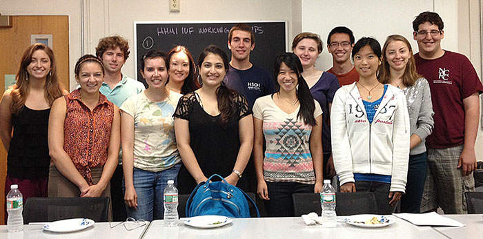
Above: 2013 HHMI Interdisciplinary Undergraduate Fellows
Sickle Cell Anemia: Functional Validation of Genetic Mutations and Human Disease
Content Creator Credits: Sarah Chapin 16
Sarahs faculty advisor: Pardis Sabeti
Tiny variations in the human genome can be significant when they impact protein function. Experimental functional analysis of these variations helps illuminate the biological effects of genetic mutations and provides further insight into the genetic basis of disease. In the case of sickle-cell disease, functional analysis allows for the determination that a genetic mutation alters the structure of hemoglobin, distorting the shape of red blood cells and hindering oxygen transport throughout the body. However, other mutations may be located in non-coding regions of the genome, and functional analysis can show that these mutations have different biological effects, such as altering the amount of protein in the cell, rather than the structure of the protein itself. In this way, understanding the biological impact of a disease-associated mutation can provide insight into the underlying physiological mechanism of the disease.
Evolutions Creative Solutions: Two sources of Protein Diversity
Content Creator Credits: Natalie Maria 16 & Viviana Maymi 16
Natalie’s faculty advisor: Michael Springer
Viviana’s faculty advisor: John Calarco
As macromolecules that are found in every living organism, proteins are essential for life, as we know it. They perform a strikingly broad array of functions that facilitate dynamic cellular activity. How can a single macromolecule have enough functional variety for it to be crucial for all forms of life? Our animation explains two important sources of proteomic diversity: gene duplication and alternative splicing. These processes increase the diversity of protein products arising from genes.
Mechanisms of Human Learning
Content Creator Credits: Timothy Hopper 14 & Rachel Zsido 14
Timothy’s faculty advisor: Robert Stickgold
Rachel’s faculty advisor: Mohammed Milad
This animation is an exploration of the amazing learning mechanisms of the human brain. Rachel studies the neural mechanisms of fear conditioning, hoping to better understand the specific brain mechanisms that govern this process. Likewise, Timothy is intrigued by the neural mechanisms underlying selective memory consolidation. Their research illustrates the truly remarkable capacity of the brain to adapt to its changing environment.
NexGen DNA Sequencing: A Look Into Microscopic Worlds
Content Creator Credits: Mia Bertalan 16 & May Yang 15
Mia’s faculty advisor: Lauren O'Connell
May’s faculty advisor: Max Nibert
Improvements in sequencing technology have lowered costs, improved efficiency, and increased the scope of sequencing applications. One advancement is Next Generation sequencing, or NexGen, which allows simultaneous identification of hundreds to thousands of unique sequences in a single sample. Utilizing this method, we can study the diversity present in microcommunities composed of thousands of different bacteria, fungi, and other microorganisms.
In Vitro Manipulation of Stem Cells for Regenerative Medicine
Content Creator Credits: Jenny Makovkina 16 & Max Zacher 15
Jenny’s faculty advisor: Tracy Young-Pearse
Max’s faculty advisor: Andrew Brack
Stem cells are a source of regeneration for many of the body's tissues, and are critical for the development and maintenance of multicellular organisms. Scientists have just recently begun to better understand the many roles stem cells play in the body, and our ever-increasing ability to control and reprogram stem cells holds promise for the future of regenerative medicine. This animation will introduce you to the basics of what makes a cell a stem cell, and will then guide you through the journey a fibroblast cell makes to first become an induced pluripotent stem cell and finally a functional muscle cell.
Neurospora crassa: The forgotten model system
Content Creator Credits: Ethan Addicott 14
Ethan’s faculty advisor: Anne Pringle
While Neurospora crassa was the model system of choice for Tatum and Beadle in their 1958 Nobel Prize winning research, this highly valuable system has since fallen out of the spotlight. Using the formaldehyde oxidation pathway as an example, we show how the filamentous fungus Neurospora crassa provides us with many tools to understand this and other important biological pathways.
CRISPR: DNAs Text Editor
Content Creator Credits: Rayhnuma Ahmed 14 & George Lok 16
Rayhnuma’s faculty advisor: Suneet Agarwal
George’s faculty advisor: Steve Hyman
Advances in genome editing allow us to manipulate a genome, whether it be to remove, insert, or replace DNA, using site-specific nucleases. This animation discusses the mechanism behind the latest editing tool, CRISPR, and walks through two applications of CRISPR. This exciting technology is simplifying genetic engineering by making it more rapid and accurate.
Visualizing Neuronal Activity via Calcium Signaling
Content Creator Credits: Julia Nguyen 14 & Vivian Yeong 14
Julia’s faculty advisor: Yun Zhang
Vivian’s faculty advisor: Pam Silver
Numerous biological phenomena occur at the subcellular level beyond the resolution of the human eye. So how are scientists able to visualize important cellular and molecular interactions? In this animation, we use calcium imaging as an example of molecular technology that allows us to both observe neuronal activity and associate complex behaviors with physiological states. Moreover, we bring to attention the importance of high-resolution visual and quantitative techniques in understanding the pathophysiology of various human diseases, such as Schizophrenia, Alzheimer’s disease, and Huntington’s disease.
The project was funded by an award from the Howard Hughes Medical Institute.
Conception and content of the animations by the Interdisciplinary Undergraduate Fellows. Student mentoring by Jacqueline M. Brooks. Animations by Matthew Bohan.